In a move that could redefine clinical orthopaedic practice, two new open-access papers reveal that a standard smartphone can rival high-end laboratory equipment, and even sharpen surgeons’ own eyes, when measuring active shoulder range of motion. The research, born of a collaboration between the Australian Research Council Training Centre for Joint Biomechanics and medical device giant Zimmer Biomet, promises to make shoulder assessments more objective, accessible and consistent.
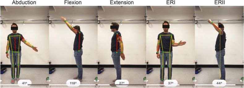
The first study pits a 2D-pose estimation algorithm, running on a smartphone camera, against the gold-standard three-dimensional motion capture systems typically confined to specialised biomechanics labs. Researchers asked healthy volunteers and patients with shoulder complaints to perform common movements, flexion, abduction and external rotation, while simultaneously recording data with both methods. The results were surprising. Smartphone estimates aligned closely with 3D motion capture for most movements, often within a few degrees. When discrepancies arose, they could be traced to slight differences in how anatomical frames were defined and to subtle thoracic motions that fall outside pure shoulder movement. By mapping these nuances, the authors have laid the groundwork for clinicians to interpret smartphone-derived angles reliably, bridging the divide between lab precision and the variability of real-world care.
Just as compelling, the second paper examines how smartphone measurements stack up against surgeons’ own visual estimates in a busy clinic setting. Orthopaedic surgeons and physiotherapists routinely gauge a patient’s shoulder mobility by eye, a practice that can vary widely from one practitioner to the next. In this study, experienced clinicians recorded their assessment of each patient’s shoulder excursion while a smartphone captured the same motion. Analysis showed that 2D-pose estimates matched surgeon judgments within five degrees for most movements. Beyond that close agreement, however, smartphone data offered a finer resolution, flagging subtle asymmetries that might escape even a trained eye. The technology thus offers a path to reduce inter-observer variation, ensuring that patients receive consistent evaluations no matter which specialist they see.
Taken together, these studies signal a shift toward democratising biomechanical analysis. No longer must clinicians choose between the high cost and limited availability of motion-capture laboratories and the subjectivity of visual estimation. With a device that most practitioners already own, they can gather precise, repeatable data in real time, enhancing decision-making throughout rehabilitation and post-operative monitoring.
These breakthroughs result from the combined expertise of the ARC Training Centre for Joint Biomechanics and Zimmer Biomet. ARC ITTC JB researchers involved in the publication include Dr Wolbert van den Hoorn, Associate Professor Kenneth Cutbush, Professor Ashish Gupta, Professor Graham Kerr, Dr Freek Hollman, Dr Roberto Pereyon Valero, Dr Max Lavaill and PhD candidate François Bruyer-Monteleone.