Pioneering the Future of Orthopaedics

A/Prof Saulo Martelli

Pioneering the future of Orthopaedics.

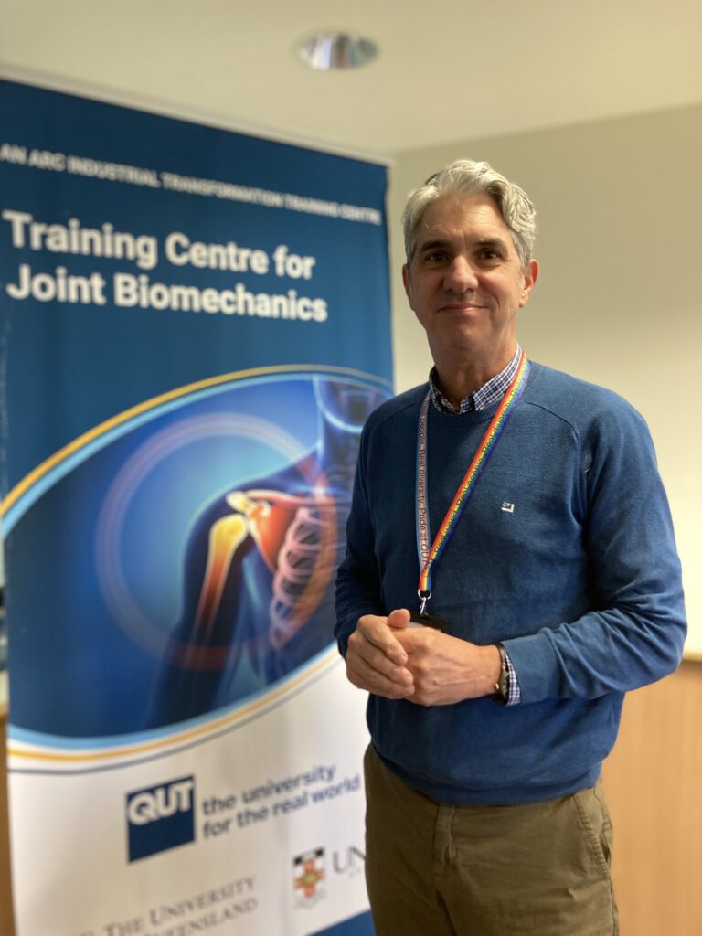
Associate Professor Saulo Martelli stands at the forefront of research that is reshaping how we understand and treat joint conditions. As Program 2 Lead at the Australian Research Council Industrial Training Centre for Joint Biomechanics, he has been guiding a bold initiative that fuses robotics, ultrasound, and biomechanics into a single, powerful vision. It is a vision that is as ambitious as it is practical: to transform cutting‑edge engineering into better surgical procedures, safer implants, and more effective rehabilitation for patients. 

Bringing Robots and Ultrasound into the Lab.

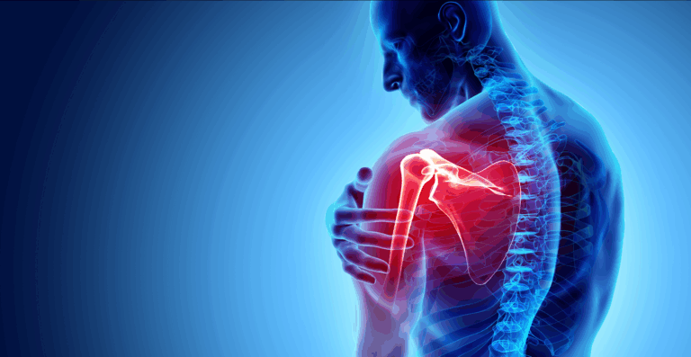
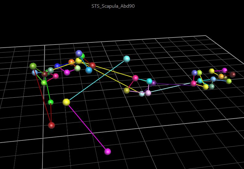
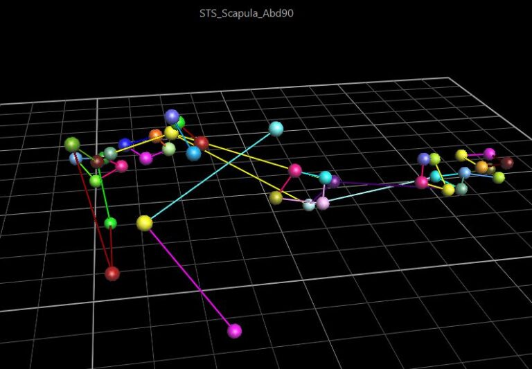
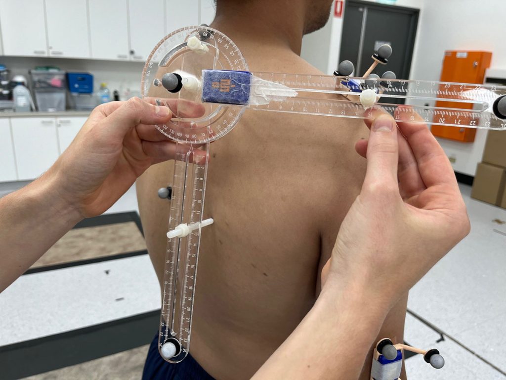
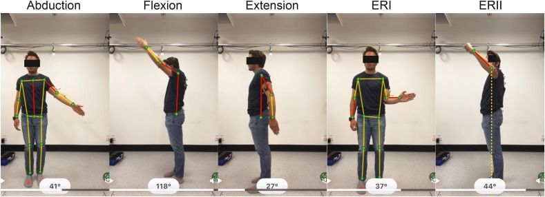
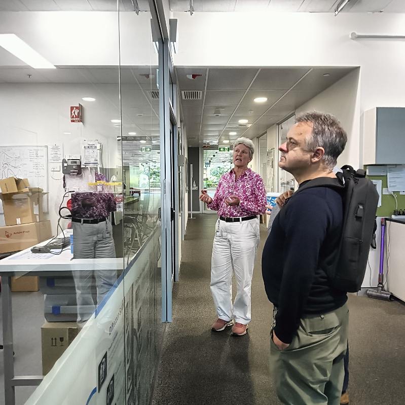
Program 2 is devoted to Robot Assisted Testing and Surgery, a field that demands both technical mastery and clinical insight. Joints like the shoulder endure complex, three‑dimensional forces that shift with every movement, making them a challenge to study and replicate. Martelli’s approach harnesses robotic systems capable of mimicking these changing loads, paired with high‑frequency ultrasound ADVANCED imaging that can detect microscopic changes in bone and implant structures. This unique combination allows his team to capture the intricate interplay between movement and stability in ways that were previously out of reach.

Transforming Shoulder Surgery with Data‑Driven Precision.

The insights gathered in the lab are more than academic data points, they are stepping stones toward tangible surgical improvements. By understanding precisely how an implant behaves under real‑world conditions, Martelli’s work helps guide surgeons toward more accurate shoulder replacements. These advances promise to increase the precision of surgical interventions, reduce recovery times, and improve long‑term joint function. In parallel, the lab’s rigorous testing protocols ensure that implants are evaluated under the most realistic and demanding conditions before ever reaching a patient.

Cracking the Code of Virtual Trials.

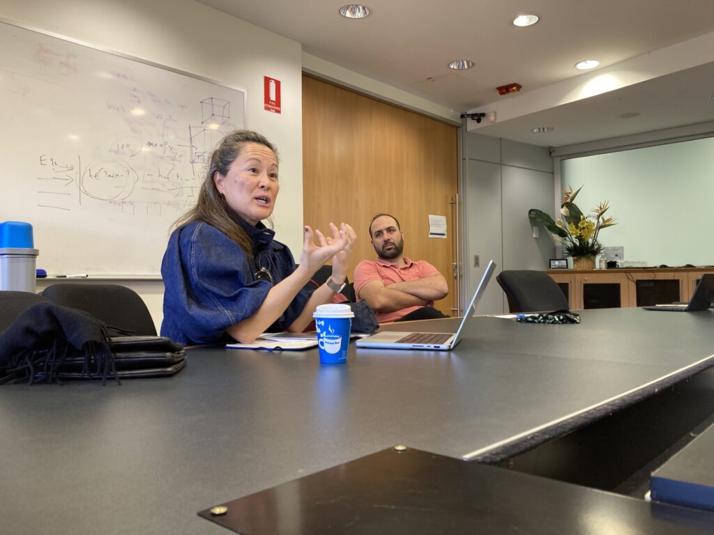
One of Martelli’s most notable breakthroughs emerged from a Zimmer Biomet collaboration with colleagues Christine Mueri, Lukas Connolly, Jeff E. Bischoff, and Philippe Favre. The team tackled a key limitation in in silico clinical trials: computer simulations of patient outcomes. Traditional high‑fidelity models can take over an hour to run for a single case, are extremely complex in their technicality and require considerable expertise to operate.  Previously impossible in surgical planning, the ability to inform surgeons if an implant will be stable months after surgery  is becoming possible through developing a surrogate model: a fast, predictive algorithm trained on previous simulation results that delivers results in milliseconds while retaining exceptional accuracy. This leap in speed allows vastly more virtual patients to be modelled in less time, enriching datasets for implant evaluation, personalised surgical planning, and rehabilitation strategies.

The Power of Collaboration

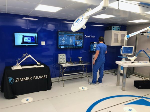
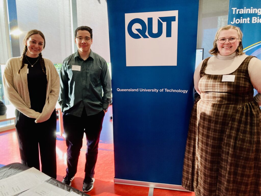
Central to Program 2’s success is its close partnership with industry, particularly Zimmer Biomet. Through the ARC Training Centre’s Impact Pathway, academic innovation collaborates with real‑world production expertise to improve diagnostic response and patient outcomes.

Taking the Vision to the World Stage

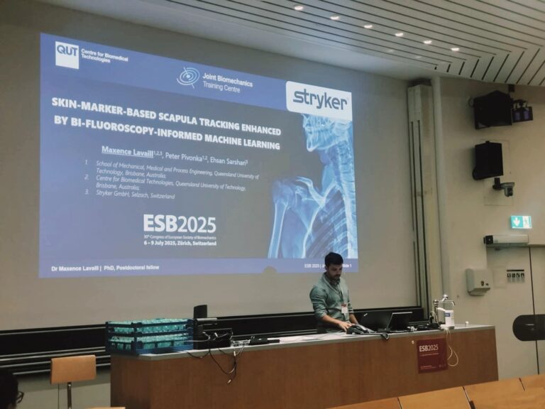
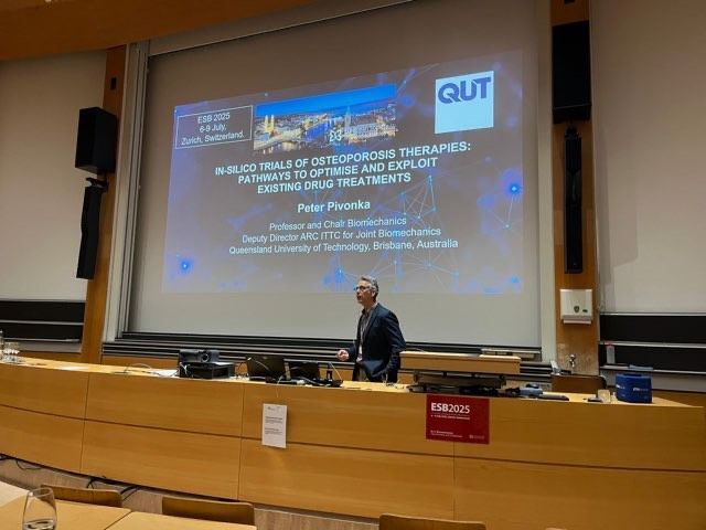
Over the coming year, Martelli’s work will be presented at major international conferences, including the ISTA 36th Annual Congress in Rome, the MDIC Symposium on Computational Modeling & Simulation in Maryland, the MDIC Annual Patient Summit, and the 9th Asian Pacific Congress on Computational Mechanics in Brisbane. These events will showcase the potential of combining robotics, ultrasound, and computational modelling to revolutionise how joint surgery is planned, executed, and assessed.

A Data‑Driven Future for Joint Care

The story of A/Prof Saulo Martelli’s Program 2 research is one of integration, bringing together engineering precision, clinical needs, and technological innovation to redefine standards in orthopaedics. By closing the gap between laboratory research and patient care, Martelli and his collaborators are charting a course toward surgeries guided by rich, real‑time data and supported by rigorous pre‑clinical validation. It is a vision in which each patient’s journey to restored mobility is shaped by science, driven by technology, and delivered with precision.

Health Sensing Technology Research Expands Into New ITTC

Prof Graham Kerr

Professor Graham Kerr to Help Lead ARC’s Push in Health Sensing.

Australia’s rehabilitation science is entering a new phase: wearable, data‑rich, and designed to work where patients actually live and recover. An important contributor to this shift is Professor Graham Kerr, Chief Investigator and Program 4 Lead at the Australian Research Council (ARC) Training Centre for Joint Biomechanics. His program specialises in in vivo assessment of upper limb movements, physiology, and rehabilitation, and has built a reputation for research that helps translate biomechanics into practical tools for clinicians and patients. Now, Prof Kerr will also serve as a Chief Investigator for the new ARC Industrial Transformation Training Centre for Transformative Health Sensing Technologies, led by the University of Melbourne, extending this trajectory into an effort focused on next‑generation wearables and sensing platforms.

Aligned missions, expanded reach.

The Joint Biomechanics Centre’s Program 4 is focused on measuring what matters: the quality, symmetry, and control of upper limb movement during real tasks, alongside the physiological signals that shape recovery. That core competency, capturing accurate human movement in the real world, maps directly onto the new Centre’s goal to build wearable devices that can continuously sense health, flag risks earlier, and personalise rehabilitation. Where Program 4 has focused expertise in upper limb function, the Transformative Health Sensing Technologies Centre will expand similar themes across a wider range of devices, contexts, and clinical pathways.

Partnerships turning research into devices.

Program 4’s approach is specifically translational. Partnering with Logemas, the team has advanced research and development around the Vicon Vero gait analysis system, high‑fidelity motion capture that provides the data needed to validate and benchmark new sensing methods. With Zimmer Biomet, the program is progressing wearable sensors and technology that can move seamlessly from clinic to home, supporting therapy adherence and ongoing monitoring between appointments.

Guided by Prof Kerr, Program 4 has also embarked on developing wearable devices built on Inertial Measurement Units (IMUs) to monitor upper limb movement in both clinical and home environments as a rehabilitation tool. IMUs offer a practical route to continuous, real‑world data without the constraints of traditional lab‑based systems. PhD Candidate Arthur Fabre is advancing this innovation pipeline with backing from Centre partners, ensuring that hardware, algorithms, and clinical validation advance together rather than in isolation.


A national investment in transformative Biotech.

ARC Industrial Transformation Training Centres are designed to do something Australia has historically struggled with: turn world‑class research into world‑class products that reshape healthcare delivery. In health sensing and rehabilitation, that transformation is already visible. Motion analysis that once required a lab is informing algorithms worn on the arm. Sensors that once delivered snapshots now stream continuous signals that map recovery in real time. And cross‑sector teams, engineers, clinicians, designers, and industry are building devices that develop grow scientific innovation and offer scalable solutions for real‑world needs.

The impact extends beyond single conditions or patient groups. Continuous, high‑quality sensing enables proactive models of care, reduces preventable hospital visits, and helps clinicians allocate attention where it will matter most. It builds a workforce fluent in both science and product, and it sets the stage for made‑in‑Australia health technologies that compete globally.

Collaboration powering shoulder biomechanics

Collaboration Powering Shoulder Biomechanics

The ARC Training Centre for Joint Biomechanics in Brisbane is building something innovative: a pipeline where cutting‑edge motion capture meets patient‑relevant science. Since 2021, Logemas, a motion-capture integrator and life sciences equipment specialist, has been an industry partner to the centre: a major collaboration between QUT, UNSW and UQ funded by the Australian Government and industry partners through the ARC Industrial Transformation Training Centre Program. That partnership has become the quiet engine behind a string of advances led by Chief Investigator Professor Graham Kerr and Dr Maxence Lavaill, translating laboratory precision into clinical promise.

 From Ground  Truth to Clinical Reality

When research asks hard questions, the answers need a benchmark. In human movement science, marker‑based motion capture remains the “ground truth” for dynamic 3D position and orientation. The centre relies on Logemas‑supplied Vicon systems to validate new and repurposed technologies, ensuring that phone‑based 2D pose methods, inertial measurement unit (IMU) pipelines and ultrasound‑derived bone tracking all stand up against the best current standard. That rigor does more than generate tidy graphs; it earns trust for tools that will ultimately guide clinical decisions.

Just as important is turning real motion into living models. The team records participant‑specific movement data to drive anatomically accurate, MRI‑informed digital models of the shoulder complex, structures that cannot be directly observed in motion, like bone and soft tissues. Rather than manipulating each joint by hand, they let captured human motion feed the model, revealing tissue behavior through lifelike simulations.

Synchronised Systems, Seamless Science

With Vicon Lock and Nexus software, the lab synchronises data across modalities without friction. Up to eight unique sync signals can be distributed, and 64 analog inputs can ingest triggers from third‑party devices to start or stop motion capture—even when no markers are recorded. The software also integrates directly with force plates, EMG, IMUs and even eye‑tracking through plugin support, making multi‑sensor experiments routine rather than heroic. That orchestration turns complex protocols into dependable workflows and gives researchers confidence that every millisecond lines up.

Prof Graham Kerr

Research Impact

Professor Graham Kerr, Chief Investigator of the Training Centre, highlights how this collaboration accelerates progress:
“Logemas has provided excellent support for all facets of motion capture including Vicon, IMUs, EMG, force plates and markerless video systems. The close synergy with Logemas has ensured efficient data capture and processing across several projects.”

For Dr Maxence Lavaill, the collaboration was formative: “Yes, it definitely was critical! Without support from Logemas, I could not have done my PhD. Half of my PhD studies was based on experimental measurements undertaken in the motion capture lab. Logemas trained me from the start of my research and supported me throughout”

Dr Maxence Lavaill

Turning Capability Into Outcomes 

At QUT’s Kelvin Grove motion capture lab (Q block), Lavaill recorded healthy participants performing basic, clinical and daily‑life shoulder motions to build a database that drives innovative MRI‑based musculoskeletal models. Surface EMG via Cometa both powers and validates these models, matching muscle activations to kinematics for a more faithful picture of shoulder function. More recently, the team used Vicon to establish ground‑truth position and motion in a cadaveric study, comparing those results with ultrasound‑based bony measurements. That kind of cross‑modality validation helps new clinical tools mature faster.

Industry impact is already visible. With Logemas technology in hand, the centre secured external collaborations and funding, validating a shoulder pose tracking algorithm from Zimmer Biomet against Vicon 3D motion capture. When companies bring ideas and labs bring proof, patients benefit sooner.

Programs, People and a Growing Ecosystem

From the outset, the centre organised its work across four programs. Logemas technology has been fundamental to Program 1, In‑Silico Upper Extremity Modelling and Simulation, and also to Program 4, supporting translation‑focused studies like clinical assessments and cadaveric validation. The other streams: Program 2 (Robot Assisted Testing and Surgery, led by A/Prof Saulo Martelli) and Program 3 (Optimised Tissue‑Engineered Scaffolds, led by Prof Justin Cooper‑White), round out a comprehensive agenda that spans devices, biology and computation.

The skills this partnership fosters are durable. “Absolutely,” Kerr says when asked if he’ll continue using Logemas beyond centre projects. Lavaill adds that the competencies are “highly sellable,” noting plans for a portable motion capture system to support biomechanical and cadaveric experiments, patient outcome assessments, and undergraduate teaching.

Smartphones prove surprising in Shoulder Motion Assessment in Centre research.

In a move that could redefine clinical orthopaedic practice, two new open-access papers reveal that a standard smartphone can rival high-end laboratory equipment, and even sharpen surgeons’ own eyes, when measuring active shoulder range of motion. The research, born of a collaboration between the Australian Research Council Training Centre for Joint Biomechanics and medical device giant Zimmer Biomet, promises to make shoulder assessments more objective, accessible and consistent.

The first study pits a 2D-pose estimation algorithm, running on a smartphone camera, against the gold-standard three-dimensional motion capture systems typically confined to specialised biomechanics labs. Researchers asked healthy volunteers and patients with shoulder complaints to perform common movements, flexion, abduction and external rotation, while simultaneously recording data with both methods. The results were surprising. Smartphone estimates aligned closely with 3D motion capture for most movements, often within a few degrees. When discrepancies arose, they could be traced to slight differences in how anatomical frames were defined and to subtle thoracic motions that fall outside pure shoulder movement. By mapping these nuances, the authors have laid the groundwork for clinicians to interpret smartphone-derived angles reliably, bridging the divide between lab precision and the variability of real-world care.

Just as compelling, the second paper examines how smartphone measurements stack up against surgeons’ own visual estimates in a busy clinic setting. Orthopaedic surgeons and physiotherapists routinely gauge a patient’s shoulder mobility by eye, a practice that can vary widely from one practitioner to the next. In this study, experienced clinicians recorded their assessment of each patient’s shoulder excursion while a smartphone captured the same motion. Analysis showed that 2D-pose estimates matched surgeon judgments within five degrees for most movements. Beyond that close agreement, however, smartphone data offered a finer resolution, flagging subtle asymmetries that might escape even a trained eye. The technology thus offers a path to reduce inter-observer variation, ensuring that patients receive consistent evaluations no matter which specialist they see.

Taken together, these studies signal a shift toward democratising biomechanical analysis. No longer must clinicians choose between the high cost and limited availability of motion-capture laboratories and the subjectivity of visual estimation. With a device that most practitioners already own, they can gather precise, repeatable data in real time, enhancing decision-making throughout rehabilitation and post-operative monitoring.

These breakthroughs result from the combined expertise of the ARC Training Centre for Joint Biomechanics and Zimmer Biomet. ARC ITTC JB researchers involved in the publication include Dr Wolbert van den Hoorn, Associate Professor Kenneth Cutbush, Professor Ashish Gupta, Professor Graham Kerr,  Dr Freek Hollman, Dr Roberto Pereyon Valero, Dr Max Lavaill and PhD candidate François Bruyer-Monteleone.

Translational Research Collaboration Strengthens Australia’s MedTech Innovation Ecosystem

Stryker Delegation Visits ARC ITTC Joint Biomechanics to Advance Additive Manufacturing and Joint Biomechanics

The ARC Training Centre for Joint Biomechanics welcomed a senior delegation from medical technology leader Stryker on Aug 6 to Queensland University of Technology (QUT). Their visit celebrated five years of impactful partnership and showcased the power of translational research to transform orthopaedic innovation. Key figures from Stryker’s Additive Manufacturing workstream Naomi Murray, Catherine Madigan, and Conor Kelleher joined Centre researchers for two days of immersive discussions.

Fostering Dialogue Between Industry and Academia

The agenda kicked off with a roundtable with Stryker featuring ARC ITTC JB Centre Director Professor Yuantong Gu alongside PhD candidate Ahmed Sewify and Centre Manager Rosalee Armitage. Together, they examined emerging trends in joint biomechanics and the future of biomedical research at QUT. Their exchange underscored how academic research and industry expertise can coalesce to address clinical challenges.

A series of presentations by Associate Professor Saulo Martelli and Professor Graham Kerr highlighted QUT’s state-of-the-art gait laboratory. Delegates observed real-time motion capture and pressure-sensing demonstrations that model patient-specific movement patterns. Conversations around smart implants, advanced gait analysis and novel pressure sensing technologies sparked inspired conversations for integrating additive manufacturing into next-generation orthopaedic devices.

Touring Cutting-Edge Microscopy Facilities

Under the guidance of Professor Charlotte Allen, the group explored QUT’s Central Analytical Research Facility (CARF). The tour showcased electron microscopy suites alongside bright-field (phase contrast and DIC) and multi-channel fluorescence microscopes. Delegates also glimpsed live-cell imaging workflows and confocal laser scanning systems, capabilities that are critical for characterizing biomaterials at nano- to micro-scales.

Since 2020, Stryker, QUT and the ARC Training Centre have forged a research alliance focused on orthopaedic research, especially within shoulder mechanobiology, personalised device development, and computational modelling. This collaboration has supported numerous student projects, and prototyping efforts, culminating in patent filings and pre-clinical studies that aim to bring smarter implants and surgical tools to market.

Advancing Shoulder Musculoskeletal Modelling

Building on this strong foundation, Centre postdoctoral fellow Dr Maxence Lavaill has secured an Advance Queensland Industry Research Fellowship (AQIRF) to investigate soft-tissue mechanics in the shoulder. His work employs patient-specific computational models to predict implant performance under physiological loads, laying the groundwork for bespoke surgical solutions that could reduce recovery times and improve long-term outcomes.

The week’s events reaffirmed the critical role of translational research collaboration. By combining QUT’s fundamental biomechanics expertise with Stryker’s commercial insights and manufacturing prowess, the partnership is bridging the gap between laboratory discovery and clinical deployment. This synergy not only accelerates product development but also strengthens Australia’s position as a global medtech innovation hub.

Looking Ahead: New Frontiers in Biomechanics

As the ARC Training Centre for Joint Biomechanics and Stryker look toward their sixth year of partnership, they plan to expand into smart biomaterials, in-silico trials, and AI-driven design workflows. Upcoming initiatives include collaborative workshops on machine-learning algorithms for implant optimization and joint user studies involving patients and surgeons. Together, they are poised to explore uncharted territory in #biomechanics and deliver innovative solutions that improve lives.

This week’s visit not only celebrated past achievements but also set the stage for future breakthroughs. With shared vision, mutual trust, and unwavering commitment, the collaboration between the ARC Training Centre and Stryker continues to push the boundaries of medical technology and reinforces Australia’s leadership in the global medtech ecosystem.

BIOTech Futures Challenge 2025 Ignites STEM Passion Across Australia 

Australia’s leading science and innovation program for high school students is once again driving change in classrooms and research labs alike. The BIOTech Futures Challenge 2025, led nationally by the University of Sydney, is equipping the next generation with the tools to tackle some of the world’s most complex issues from biomedical breakthroughs to climate resilience. 

A flagship initiative designed to reimagine the role of STEM in education, BIOTech Futures connects high school students from urban, regional and remote communities with academic mentors from universities and research centres. Since its inception, over 2,000 students from 80 schools have taken part, producing more than 400 research projects guided by 300 academic experts. 

In Queensland, the Challenge is proudly hosted by the ARC Training Centre for Joint Biomechanics at the Queensland University of Technology (QUT). Since 2021, the Centre has led the state chapter with a mission to ignite STEM curiosity and drive student innovation. Through this platform, staff, including chief investigators, research fellows, PhD candidates and industry-established alumni, mentor student teams for six weeks, helping them address real-world problems with creative, science-driven solutions.

Students will showcase their work, including posters, short answer responses, and prototypes, at a symposium at QUT on October 9. Top projects will proceed to the national symposium at the University of Sydney on October 24, representing Queensland on a national stage. 

Projects can span areas such as AI-driven diagnostics, sustainable materials for healthcare, and ethical design of emerging technologies. But behind the innovation lies a national concern: experts are warning of a steady decline in domestic STEM enrolments. Data from the Federal Government’s Tertiary Collection of Student Information (TCSI) shows a 3.1% drop in 2022, followed by a 1.4% drop in 2023, with enrolments in natural and physical sciences falling by 11%. 

STEM education is more than academic, it’s foundational to Australia’s prosperity. With 75% of jobs in the fastest-growing industries requiring STEM skills, the nation’s ability to remain competitive hinges on cultivating a workforce equipped for technological change. Experts warn this trend could lead to a Brain Drought, with far-reaching impacts on workforce development and economic competitiveness. 

By connecting students with mentors and research environments, the Training Centre for Joint Biomechanics is actively working to reverse this trend. Its efforts are part of a broader movement to make STEM accessible, exciting, and relevant. STEM plays a crucial role in fostering critical thinking, creativity, and problem-solving: skills essential for navigating the challenges of the 21st century. 

“Ambitious students have the ability to develop innovative ideas and technologies that can change the world,” said Prof. Hala Zreiqat AM, founder and director of BIOTech Futures. This Challenge rewards those with the persistence and tenacity to develop their ideas.” 

Dr. Victoria Camilieri-Asch, mentor to last year’s winning QLD team, believes in the power of immersive STEM engagement: It has impact if students can see our labs and research environments. Sharing our own views and experiences can really enrich students’ understanding through real-life applications.” 

As Australia faces critical skills shortages in science and technology, BIOTech Futures continues to prove that investment in young minds today shapes the discoveries of tomorrow. 

The Road to Impactful Collaborations at the European Society of Biomechanics Congress

Dr Maxence Lavaill

Advancing Non-Invasive Scapular Tracking

In July 2025, the Australian Research Council Industrial Transformation Training Centre for Joint Biomechanics underscored its dedication to tackling critical challenges in musculoskeletal research at the European Society of Biomechanics Congress in Zurich. Dr Maxence Lavaill delivered an impactful presentation entitled “Skin-Marker-Based Scapula Tracking Enhanced by Bi-Fluoroscopy-Informed Machine Learning,” confronting one of shoulder biomechanics’ longest-standing obstacles: accurately measuring the scapula’s complex three-dimensional motion through layers of soft tissue.

Conventional videogrammetry setups using skin markers can err by as much as 80 mm in position and 25° in orientation, while dynamic bi-fluoroscopy bypasses soft tissues but remains prohibitively expensive and reliant on ionising radiation. Dr Lavaill’s work leverages a recently published dataset in which twenty healthy volunteers performed seven shoulder movements—coronal and scapular abduction, forward flexion, two internal rotations, and two external rotations—while ten thoracic, scapular, upper arm, and forearm landmarks were tracked by a ten-camera Vicon system and five scapular landmarks were captured at 100 Hz via a custom bi-fluoroscopy rig.
 
Employing demographics (sex, age, height, weight), motion type, shoulder side and a sliding window of three-dimensional marker coordinates across four preceding frames, the study constructed 105 predictors to anticipate 15 ground-truth fluoroscopic landmark coordinates over 101,936 motion frames. Sixteen participants’ data trained ensemble-bagged-trees regression models in MATLAB’s Regression Learner App, with the remaining four reserved for validation. Predicted marker trajectories were then filtered to ensure temporal continuity and benchmarked against raw skin-marker errors.
Dr Lavaill at the ESB Congress in Zurich
Demonstrable Improvements and Clinical Promise
The machine-learning models consistently diminished tracking errors, achieving landmark accuracies between 4 and 14 mm—up to a 70 percent reduction compared to conventional skin markers. Scapular orientation errors fell from an average of [− 0.4, 10.1, − 10.2]° using Vicon landmarks to [− 2.7, 4.1, 4.5]° after ML correction, aligning performance far closer to fluoroscopy’s precision without associated drawbacks. As Dr Lavaill remarked, “This research tackles one of shoulder biomechanics’ trickiest challenges—accurately tracking scapular motion non-invasively. By integrating skin-marker data with the precision of bi-planar fluoroscopy through machine learning, we’re pushing boundaries in motion analysis, sports science, and clinical diagnostics.”
Expanding Computational Trials in Bone Therapies
Deputy Centre Director Professor Peter Pivonka complemented this work with his lecture, “In-Silico Trials of Osteoporosis Therapies: Pathways to Optimise and Exploit Existing Drug Treatments.” He outlined how high-fidelity bone-remodelling models can simulate virtual patient cohorts to predict dosing regimens, assess long-term outcomes and refine therapeutic strategies—presenting an urgent alternative to costly early-phase clinical trials. Together, these presentations underscored the ESB Congress’s core focus on bridging experimental and computational modelling to solve biomechanical complexities.
Prof Peter Pivonka at the ESB Congress, Zurich
The Imperative of Scientific Exchange
For Dr Lavaill, engagement at international congresses is central to scientific advancement. Presenting six months of data at the Congress allowed for solicitation of peer feedback on the open-source model, and it gauged the interest from academic and industry delegates, identifying colleagues whose insights might drive subsequent software refinements. He has already earmarked the International Society of Biomechanics Congress later this year and the 2026 World Congress of Biomechanics in Canada as critical venues for wider validation, integration with wearable sensor platforms, and exploration of population-specific adaptations. However the focus on modelling as explored in the ESB Congress is the most closely aligned with Dr Lavaill’s research.
Forging Global Partnerships
Building on dialogues initiated in Zurich, Dr Lavaill will embark on collaborative studies in the United States and in Switzerland. These strategic collaborations not only deepen the ARC ITTC for Joint Biomechanics’ international footprint but also affirm its mission to translate computational and experimental innovations into tangible health solutions.

ARC Training Centre Workshop Equips Researchers to Maximise Industry Impact

The ARC Training Centre for Joint Biomechanics recently hosted a dynamic professional development workshop aimed at equipping its researchers with the tools to communicate the significance of their work well beyond academia. Titled “Capturing the Impact of Your Research,” the Brisbane-based session focused on strategies for articulating research value, improving visibility in the public domain, and fostering networks to advance career opportunities. 

Central to the event was a keynote presentation by Chris Cahill, Queensland University of Technology’s Research Engagement and Impact Coordinator. Cahill addressed the critical role of research impact in shaping career trajectories and securing future funding. He highlighted that visibility can transform projects from isolated discoveries into collaborative platforms and policy-shifting outcomes.  

“Placing research in the public sphere allows it to be accessed, applied, and extended,” Cahill said. “It changes the national funding conversation by demonstrating return on investment and opens doors to new partnerships.” 

Cahill outlined the dual role of outputs such as publications, reports, and presentations, and outcomes like findings, knowledge, and capability-building. These elements together form the foundation of a strong engagement strategy, leading to tangible and traceable impacts. He urged researchers to reflect retrospectively by asking key questions: Why was the research needed? What were the findings? Who used it? How was it applied? And what changed as a result?

Complementing Cahill’s insights, HDR Career Educator Karen Cavu delivered a compelling call to action on enhancing visibility and personal branding. She cited a striking statistic: “Only 10% of academic papers are read by more than 10 people outside the immediate research community. That means 90% of critical knowledge remains hidden from public view.” 

Cavu challenged attendees to rethink how they showcase their work. Her recommendations included engaging in public discussions, celebrating and supporting professional networks, publishing and sharing research broadly, updating university and conference biographies, and leveraging awards and recognitions to build professional credibility. 

Interactive segments allowed attendees to workshop their own visibility strategies, with many praising the relevance of the advice. Workshop attendee and Centre Post-Doc Fellow Ahmed Sewify commented “As a recent PhD graduate, I found Karen and Chris’s workshop incredibly timely and insightful. They clearly outlined the differences in how impact should be communicated in academia versus industry. Their practical advice helped me rethink how I frame my work for broader audiences, and I left feeling much more confident in presenting both my research and personal narrative moving forward.” 

The workshop marks an important step in the ARC Training Centre’s mission to foster not only scientific excellence but also societal impact. As joint health and biomechanics face increasingly complex challenges, preparing researchers to be skilled communicators and strategic collaborators is vital to translating research into real-world change. 

This event underscores the Centre’s commitment to shaping future leaders who have tangible impacts on the industries they are seeking to transform.

Rethinking Osteoporosis: A Digital Leap Forward from ESB 2025 

At the recent European Society of Biomechanics (ESB) Congress held at ETH Zurich, leaders in biomedical technology gathered to explore how artificial intelligence is reshaping the future of healthcare. Among the perspective talks was a fascinating presentation by Professor Peter Pivonka, Deputy Centre Director of the ARC Training Centre for Joint Biomechanics. 

Prof Pivonka’s talk “In-Silico Trials Of Osteoporosis Therapies: Pathways To Optimise And Exploit Existing Drug Treatments” detailed emerging approaches called in-silico trials. These are computer simulations that mimic how bones change over time and how treatments behave inside the body. These trials offer doctors and researchers a way to “test” therapies virtually before applying them in the real world. 

This method could have a real impact on how osteoporosis is diagnosed and treated. Traditionally, medications like denosumab have been prescribed based on broad age-related guidelines. But Prof Pivonka’s research shows that timing matters far more than we thought, especially for women entering menopause. 

His team found that if denosumab is given too early, for example before the age of 60 or within 10 years of menopause, its efficacy may be challenged. That insight could help refine treatment plans and avoid unnecessary costs or side effects from poorly timed therapies. 

For clinicians, these virtual trials offer a glimpse into a future where treatment isn’t one-size-fits-all. Instead, healthcare providers could use simulation data to tailor osteoporosis therapies based on each patient’s unique biology, age, and health history. 

By simulating how bones degrade and rebuild, and analysing the molecular factors that drive those changes, this research helps distinguish between different types of osteoporosis—like postmenopausal versus age-related. That distinction is crucial, as it guides doctors towards more precise prescriptions and better patient outcomes. 

Prof Pivonka’s work goes beyond the lab. It’s part of a bigger movement to bring advanced computing and biomedical engineering directly into hospitals and clinics. With AI-driven models and virtual testing, researchers can identify which existing medications work best, fast-track clinical decisions, and reduce the need for large, time-consuming trials. 

This has real impact not just for specialists, but for GPs, pharmacists, and health systems navigating a growing, ageing population at risk of fractures and mobility issues. 

As biomechanics enters the AI era, Prof Pivonka’s presentation signals a shift in how medicine approaches bone health. More than just new science, it’s a new mindset: data-driven, patient-specific, and digitally powered.